Randy Lindquist
 (opens in new window)B.A., Williams College
Immunological Voyeurism: Visualizing Dendritic Cells with Transgenic Mice
presented by Michel C. Nussenzweig
Randall Lindquist is a native of New York City and a graduate of Stuyvesant High School and Williams College. He was inspired to go into medicine and science by his brother’s struggle with and cure from promyelocytic leukemia. It was therefore natural for Randy to join a laboratory that works on leukocytes and lymphoid malignancies when he came to the Rockefeller-Cornell-Sloan Kettering Tri-Institutional M.D.-Ph.D. Program.
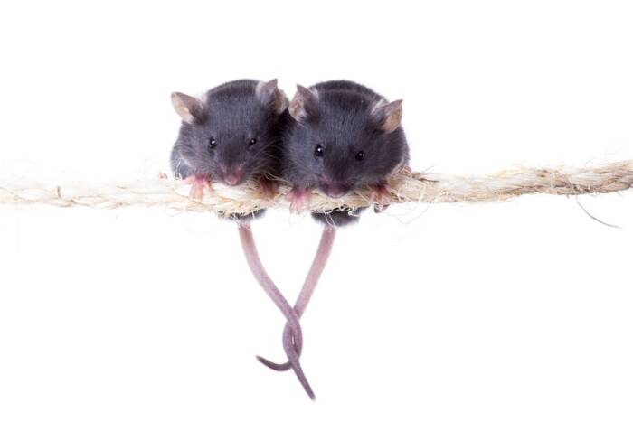
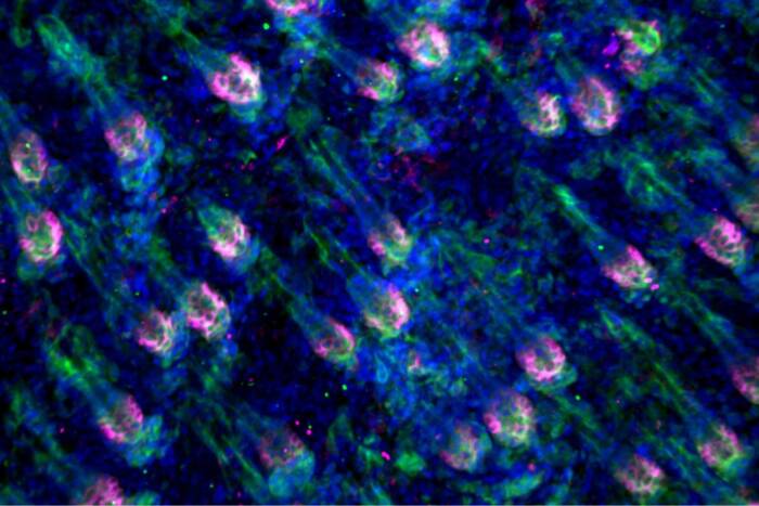
When Randy came to the laboratory he decided that he wanted to try to directly visualize dendritic cells in living tissues to learn about how they interacted with other cells, and in particular T cells, to initiate immune responses. To do this, he used a new technology called two-photon laser scanning microscopy, which allows you to make movies of cells in living mice. To see the cells, he collaborated with Hedda Wardemann to create mice in which dendritic cells glowed fluorescent green. What he saw in the microscope when he looked in living tissues is that dendritic cells exist in dense networks that look like vast colonies of sea anemones with long and constantly moving colorful extensions. He found that T cells are constantly moving through these networks in search of antigen and when they find a dendritic process with the cognate antigen, they stop to receive their marching orders and only leave after a prolonged contact-dependent conversation. Randy then collaborated with Tanja Schwickert, another Ph.D. student in the laboratory, to examine the dynamics of B cell responses in germinal centers. Randy’s work led to two first-author publications in Nature Immunology and a collaborative paper in Nature. Randy is now finishing medical school at Cornell before his next adventure in science and medicine.