Structural analysis of SpvB protein show how Salmonella bacteria hijack a cell
When Salmonella bacteria attack, they create their own compartment or vacuole in the host’s cell, in which they replicate. Now, in a cover article published this week in the journal Structure, Rockefeller University scientists show how the pathogen uses a protein called SpvB to create this compartment by breaking down the cell’s structural molecules.
 (opens in new window)
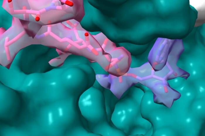
Structurally unsound. Rockefeller researchers have solved the structure of the Salmonella virulence protein SpvB (top image, in green) and shown that it works by adding a molecule to its host cell's structural proteins, thereby disrupting the cell's structural framework. SpvB shares a similar structure with other bacterial toxins, including diphtheria (bottom image, in purple) and botulism (bottom image, green).
Salmonella bacteria can wreak all kinds of intestinal havoc, from abdominal cramping to severe diarrhea. To do so, the microbe manipulates a number of the host cell’s proteins, including actin and other structural molecules normally used for assembling the cell skeleton. Erec Stebbins, associate professor and head of the Laboratory of Structural Microbiology, and his colleagues focus on a Salmonella virulence protein called SpvB — a protein in a well studied class of enzymes that includes toxins in diphtheria and pertussis (a.k.a. whooping cough). The researchers already knew that SpvB modifies actin by attaching a molecule to it, thereby disrupting the cell’s normal framework. Now, they’ve not only described the structure of SpvB for the first time, but in order to understand how it prevents actin from forming its characteristic filaments, the scientists have described the structure of SpvB-modified actin and identified the site of modification.
When they looked at the structure of SpvB, they confirmed its similarity to other members of a family of toxins called ADB-ribosyltransferases. And when they looked at the actin structures, they were able to answere a long-standing question of why modified actin is unable to form filaments. Two theories had previously been proposed: one suggested that the modification was causing actin to misfold (the “conformational change” model), and the other that something was simply being added to the molecule to prevent it from stacking (the “steric clash” model), “like taking LEGOs that would normally fit together and then putting something in there such that they could no longer stack,” Stebbins says.
Actin tends to be sticky to work with, because it clumps together in solution and precipitates out to form stringy filaments. But because the toxin prevents actin agglomeration, Stebbins was able to keep the protein in solution and crystallize two modified forms of it. And once he had the structures figured out, he could see that they hadn’t undergone large structural changes — they were the same shape as previously described actin molecules, ruling out the conformational change model. So the researchers consulted a model of actin based on images from an electron microscope, and found that the location of the SpvB modification would prevent actin molecules from stacking that way. “Very clearly, we could see, it was going to lead to packing problems,” Stebbins says.
“This is one more piece in the puzzle, at the mechanistic level, for how Salmonella is orchestrating these events to remodel the cytoskeleton.” And further down the road, he notes, knowing the structure of the molecule could lead to targeted drug design to better fight infections by Salmonella and related bacteria.