Microscopy Milestones: Celebrating 25 Years of the Bio-Imaging Resource Center
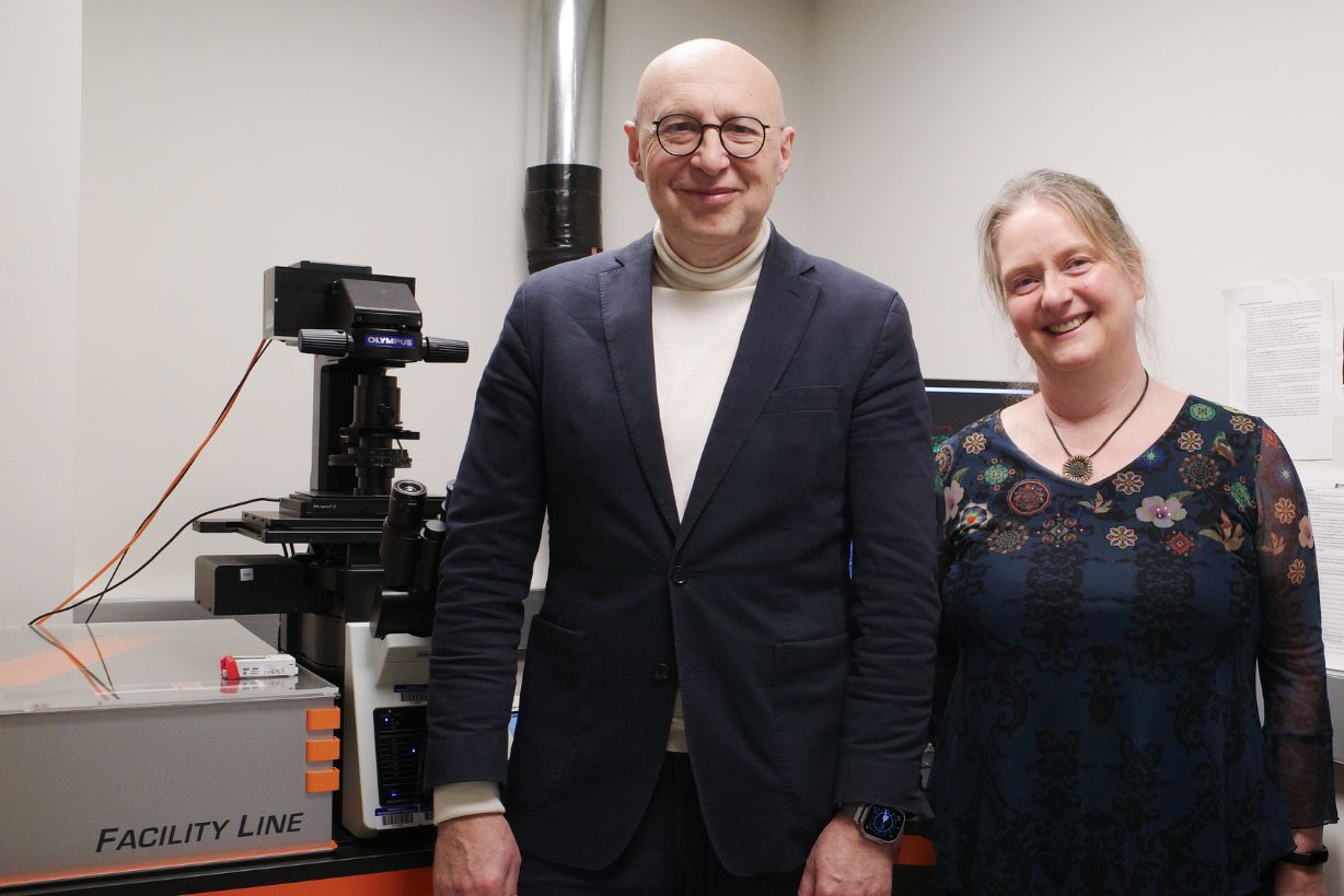
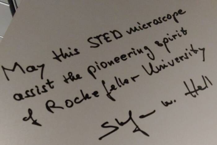
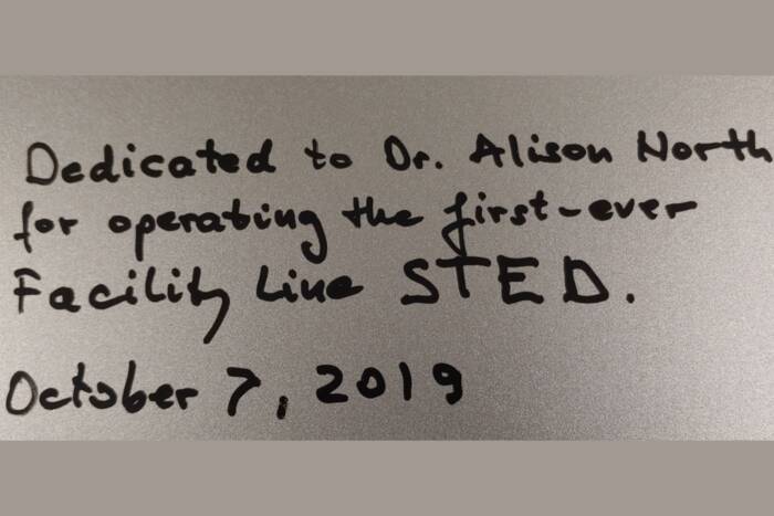
Professor Stefan Hell, inventor of STED microscopy, reunited with the 1st-in-the-world Facility Line microscope from his company, Abberior, alongside Dr. Alison North. This microscope was personally signed by Professor Hell prior to its shipping in 2019 (see his messages below).
This year, the Frits and Rita Markus Bio-Imaging Resource Center (BIRC) at Rockefeller University celebrates 25 years of advancing microscopy-intensive research. Under the leadership of Dr. Alison North, the BIRC continues to implement new imaging innovations, offering researchers cutting-edge tools and expertise to push the boundaries of discovery. Here’s a look at some recent updates:
• Super-Resolution Brilliance: The Abberior Facility Line STED system has unlocked unprecedented visualization of microscopic details, recently resolving tightly packed bacterial microcolonies in collaboration with teams from the UK and Singapore.
• Next-Generation Confocal: The new Evident FV4000 microscope, funded by HHMI, delivers unmatched sensitivity and speed, revolutionizing research capabilities on campus.
• Miltenyi Blaze: On loan until April, this upgraded light sheet microscope achieves imaging speeds up to 50x faster than previous systems, handling large sample loads such as multiple cleared mouse brains and organoids. Researchers are encouraged to test it out before it’s gone.
• BALM Lab: A new satellite laboratory dedicated to light sheet microscopy for living specimens, featuring pre-commercial systems designed around different types of biological specimens.
Super-Resolution Brilliance
The BIRC continues to have the most comprehensive range of super-resolution microscopes in the northeast region. The Abberior Facility Line STED system – the first system of its kind to be installed in the USA – continues to amaze with both its 2D- and 3D-STED modes. In 3D mode, the unparalleled axial resolution it provides, combined with custom image analysis pipelines developed by Dr. Ved Sharma, recently helped to resolve and segment tightly packed bacterial aggregates in infected cells which could not be resolved by any other light microscope here in the BIRC.
This breakthrough, published in A comparison of super-resolution microscopy techniques for imaging tightly packed microcolonies of an obligate intracellular bacterium (North, Sharma et al., 2024(opens in new window)), showcases the system’s transformative potential for microbial, as well as cell biological, research.
Next-Generation Confocal
The FV4000 confocal microscope(opens in new window) from Evident Scientific is the latest addition to the BIRC’s arsenal. Featuring ten laser lines, six advanced SilVIR detectors, and a resonant scanner, it delivers unparalleled sensitivity, dynamic range, and speed. Funded by HHMI and installed this week, this instrument is poised to transform research across campus.
Miltenyi Blaze: A High-Speed Revolution
On loan until late April, the Miltenyi Blaze light sheet microscope sets new benchmarks in speed and capacity. With imaging capabilities up to 50 times faster than previous systems, it can handle large-scale samples such as eight cleared mouse brains, 48 organoids, or even half a cleared monkey brain.
Researchers are encouraged to contact Dr. Priyam Banerjee and Dr. Maria Harreguy for hands-on testing and exploration before the system leaves campus.
BALM Lab: Pioneering Light Sheet for Living Systems
The Beckman Advanced Lightsheet Microscopy (BALM) lab, located in Bronk 102, offers groundbreaking opportunities for live specimen imaging. Led by Dr. Behzad Khajavi, the lab features pre-commercial systems such as a 3-color SCAPE microscope, inspired by Dr. Elizabeth Hillman’s design, and a Gen III Lattice Light Sheet microscope that will soon be equipped with Adaptive Optics for deeper imaging capabilities.
Both systems are ready for researchers to explore, offering fast and gentle imaging from subcellular resolution through to intact animals.
Looking Ahead
For 25 years, the BIRC has been a hub of innovation and collaboration at Rockefeller. Dr. Leslie Vosshall remarks, “The BIRC is mission critical to the Vosshall Lab – essentially every study we do relies on the instrumentation and expertise that Dr. Alison North has so steadfastly secured for RU over the past several decades. Alison is deeply committed to training the next generation of scientists in the art and science of microscopy and goes out of her way to solve problems with samples and data analysis. Alison and her team are the best!”
References:
North, A., Sharma, S., et al. (2024). A comparison of super-resolution microscopy techniques for imaging tightly packed microcolonies of an obligate intracellular bacterium. WSBM, 10(1002). https://doi.org/10.1111/jmi.13376(opens in new window)
To learn more about the Bio-Imaging Resource Center and explore its resources, visit https://www.rockefeller.edu/bioimaging.
To read more news about the Scientific Resource Centers, visit https://www.rockefeller.edu/researchsupport/news-and-announcements.
Captions for photos below:
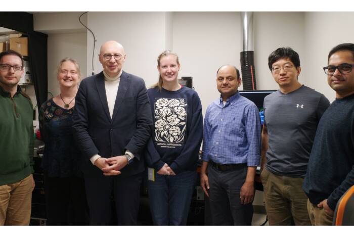
From left to right: Photo 1 shows the BIRC team with Professor Stefan Hell; Photos 2 and 3 are of messages from Professor Stefan Hell to Alison North and RU handwritten on the lid of the Abberior STED Facility Line microscope box in 2019.