The neurons in our gut help the immune system keep inflammation in check
The immune system exercises constant vigilance to protect the body from external threats—including what we eat and drink. A careful balancing act plays out as digested food travels through the intestine. Immune cells must remain alert to protect against harmful pathogens like Salmonella, but their activity also needs to be tempered since an overreaction can lead to too much inflammation and permanent tissue damage.
New research from Rockefeller University’s assistant professor Daniel Mucida, head of the Laboratory of Mucosal Immunology, shows that neurons play a role in protecting intestinal tissue from over-inflammation. Published in Cell on January 14, the findings could have treatment implications for gastrointestinal diseases such as irritable bowel syndrome.

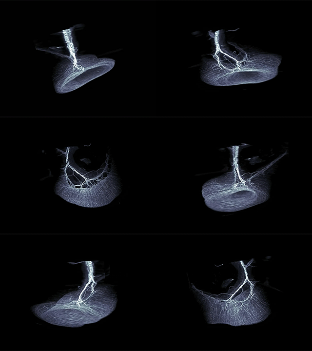
Neurons say relax: This three-dimensional view of part of a mouse intestine shows the neurons that surround tissue-protective immune cells. These neurons release norepinephrine, which instructs the immune cells to activate an anti-inflammatory response.
“Resistance to infections needs to be coupled with tolerance to the delicacy of the system,” says Mucida, who led the research together with co-first authors Ilana Gabanyi, a postdoctoral associate, and Paul Muller, a graduate student. “Our work identifies a mechanism by which neurons work with immune cells to help intestinal tissue respond to perturbations without going too far.”
Opposites react
Different populations of macrophages are among the many types of immune cells present in intestinal tissue. Lamina propria macrophages are found very close to the lining of the intestinal tube, while muscularis macrophages are in a deeper tissue layer, more distant from what passes through the intestine.
Using an imaging technique developed by Marc Tessier-Lavigne’s Laboratory of Brain Development and Repair that allows scientists to view cellular structures three-dimensionally, the researchers looked in depth at the differences between the two populations. In addition to variations in how the cells look and move, they noticed that intestinal neurons are surrounded by macrophages.
When Mucida and colleagues analyzed the genes that are expressed in the two macrophage populations, they found that lamina propria macrophages preferentially express pro-inflammatory genes. In contrast, the muscularis macrophages preferentially express anti-inflammatory genes, and these are boosted when intestinal infections occur.
“We wanted to know where this signal was coming from that induced this different response to infection,” says Mucida. “We came to the conclusion that one of the main signals seems to come from neurons, which appear in our imaging to almost be hugged by the muscularis macrophages.”
How the gut–brain axis halts inflammation
In other experiments, the scientists found that muscularis macrophages carry receptors on their surface that allow them to respond to norepinephrine, a signaling substance produced by neurons. The presence of the receptor might indicate a mechanism by which neurons signal to the immune cells to put a stop to inflammation.
The researchers also observed that the muscularis macrophages are activated within one to two hours following an infection—significantly faster than a response would take if it were completely immunological, not mediated by neurons. They believe that because these deeply embedded macrophages receive signals from neurons, they are able to respond rapidly to an infection, even though they are not in direct contact with the pathogen.
“We now have a much better picture of how the communication between neurons and macrophages in the intestine helps to prevent potential damage from inflammation,” says Mucida. “It’s plausible that a severe infection could disrupt this pathway, leading to the tissue damage and permanent gastrointestinal changes that are seen in diseases like irritable bowel syndrome. These findings could be harnessed in the future to develop treatments for such diseases.”
 Cell, online: January 14, 2016 Cell, online: January 14, 2016Neuro-immune interactions drive tissue programming in intestinal macrophages Ilana Gabanyi, Paul A. Muller, Linda Feighery, Thiago Y. Oliveira, Frederico A. Costa-Pinto and Daniel Mucida |


