Study of neurons leads scientists to re-envision vision
As we age, our eyes change shape — that’s why you see your eye doctor every year. But new research from Rockefeller University suggests that how the brain interprets visual information also changes with experience. And by studying the way in which nerve cells form connections between the eye and the brain, researchers say that some of their basic assumptions about the nature of visual development may need to be rethought.
 (opens in new window)
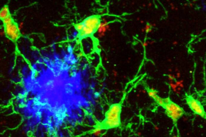
Transmitting vision. Even in adults, neurons in the brain's primary visual cortex are extremely dynamic, as researchers observed by following labeled individual neurons on a week-by-week basis. There, information is conveyed as nerve cells pass signals to one another via swellings along the axon, called boutons.
Previously, scientists had assumed that the connections between neurons in the visual pathway become fixed after a critical period early in life, but by studying neurons in monkeys, observing the same cells week to week, Charles Gilbert, head of the Laboratory of Neurobiology, now shows that they are actually changing at a high rate even into adulthood. “People had believed that the primary visual cortex, which receives information from the eye, should have fixed properties in adulthood,” says Gilbert. “What we now show is that the properties of the primary visual cortex are not fixed, and they can be continually modified to encode the information with which you become familiar throughout life.”
Using a two-photon microscope that was custom made with help from Winfried Denk, who is at the Max Plank Institute in Germany and is one of the scientists who first invented this imaging system, and a new viral labeling system developed in Gilbert’s lab, the researchers were able to examine the same neurons week after week in the primary visual cortex of adult monkeys. They identified the changes occurring in specific individual boutons, the swellings along axons, over multiple imaging sessions covering several weeks.
“Synapses, the points of contact between nerve cells at which information is exchanged, involve a presynaptic process, the axon, and a post-synaptic process, the dendrite,” says Gilbert. “The point at which the synapses occur from the axonal side is the bouton, and every bouton has at least one corresponding synapse. By following individual boutons, we could get a very good handle on the coming and going of synapses. And what we found was that there was a substantial amount of turnover week to week.”
The dynamic nature of the synapses raises the possibility that the neurons of the primary visual cortex still have a level of plasticity, or an ability to change with experience. The experiments conducted by Gilbert and colleagues, including former graduate student Dan Stettler, graduate student Homare Yamahachi and Wu Li, a research assistant professor, measured changes to the number of synapses week to week without any learning regimen or physical manipulations to the neurons.
Future experiments that look at the boutons during the learning of new shapes, or when there is interference in the exchange of information from the eye to the brain, will be able to use the results of this study as a baseline to measure how they change. “Synapses are forming and un-forming at a substantial rate, and it is against this background that experience-dependent changes take place,” says Gilbert. “We don’t yet know if all synapses are capable of undergoing change, or if only a subset has this ability. It will be interesting to find out how, as these changes occur, the brain maintains stability of some functions, even while new information is still being encoded