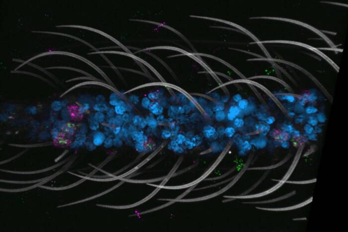
Scientists say toxins use their shape to wreak havoc on cells
Last year, Rockefeller University scientists used X-ray crystallography to determine the structure of cytolethal distending toxin (or CDT) — a widespread toxin found in diverse, disease-causing bacteria that binds to human cells, inserts a component of itself inside, then proceeds to attack the cell’s DNA. Now, these researchers have taken an even closer look at its configuration to gain a better understanding of what makes this potential carcinogen so effective.
 (opens in new window)
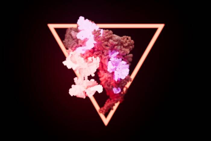
Blocking a toxin. When mammalian cells are exposed to a normal CDT toxin (green areas, top) the toxin quickly gains entry and begins attacking the cell's DNA. A mutant form of CDT which lacks a structure called the aromatic patch, however, is unable to get inside the cell (bottom).
In a paper published in PLoS Pathogens, C. Erec Stebbins, head of Rockefeller University’s Laboratory of Structural Biology, and Dragana Nesic, a research associate in the lab and the lead author, investigated two structural components of CDT. Stebbins’ prior research had shown that the molecule consists of three subunits, CdtA, CdtB, and CdtC. These subunits create a distinctive shape with two important properties: A long deep groove at the center of the molecule, and a cluster of ring-shaped amino acids, called an aromatic patch, perched at the top of the CdtA subunit. Now, Stebbins and Nesic have found that both of these features are vital not only to the molecule’s toxicity, but also its ability to bind to a cell’s surface then slip a piece of itself — the toxic CdtB subunit — inside.
The researchers mutated the CDT molecule to selectively disrupt the activity of either its groove or its aromatic patch. Then, they attached fluorescent labels to both the mutants and to intact CDT, to track and compare their ability to attach to cells and inject them with toxin. When Stebbins and Nesic subjected human cells to the fluorescently tagged CDT, they found that the cells exposed to the groove mutants glowed less than those exposed to undamaged CDT molecules. Cells subjected to the aromatic patch mutants hardly glowed at all.
“The aromatic patch mutant completely loses cell surface binding.” Stebbins says. “With this paper, we’ve confirmed that the groove and aromatic patch are the surface features that are key for binding to cells.” And the better scientists understand this genotoxin, Stebbins says, the better they’ll be able to search for molecules with similar properties that can compete with CDT, binding to a cell before the toxin can and thus preventing it from wreaking havoc.
PLoS Pathogens 1(3): November 2005(opens in new window)