Never too much of a good thing
Changing levels of a single protein can produce many different outcomes
An ongoing scientific argument surrounds the Wnt protein: Different research groups say that Wnt proteins are involved in cell differentiation, proliferation, fate determination, stem cell self-renewal and cancer. But which group is right? New research from the laboratory of Elaine Fuchs, Ph.D., shows that the answer may be that Wnt proteins can be, and are, involved in many if not all of these processes, and it is the amount of Wnt proteins that help determine what the outcome will be.
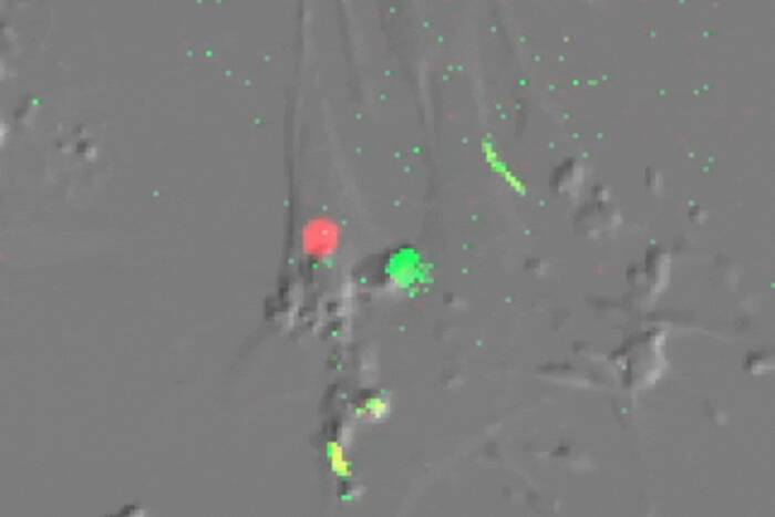
“In our previous work, we made a mouse where cells that received a Wnt signal would turn blue,” says Fuchs, Rebecca C. Lancefield Professor and head of the Laboratory of Mammalian Cell Biology and Development at Rockefeller and a Howard Hughes Medical investigator. “In that study, we had seen blue cells in the compartment of dormant (resting) skin stem cells, in their rapidly proliferating progeny that were differentiating to form the hair, and in the embryonic skin stem cells as they were choosing to become a hair follicle rather than make epidermis. When we artificially elevated Wnt signaling in the skin, the mice developed skin tumors that also had blue cells. Hence we suspected that Wnt signaling was involved in all of these different stages.”
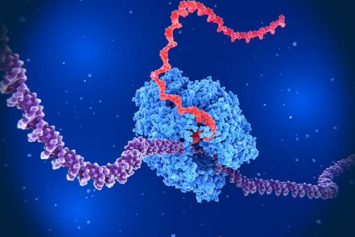
The major result of an external Wnt signal is that a protein called beta-catenin, located inside the cells, becomes stabilized and moves into the cell nucleus. Once there, beta-catenin can participate in turning on other genes. In research conducted primarily by two of her postdoctoral fellows, William Lowry and Cedric Blanpain, Fuchs and her colleagues looked at beta-catenin in the stem cell compartment found in hair follicles.
“Hair follicles undergo periodic cycles of growth, destruction and rest. In order to activate a new round of hair growth, stem cells in this compartment must be activated,” explains Fuchs. Exactly how stem cells become activated was unknown. Fuchs and her team discovered that slightly elevated levels of beta-catenin cause the stem cells to be activated prematurely, pushing them to divide and start the new hair cycle. Once activated, the stem cells return to a resting state.
“In the skin, the dose of stabilized beta-catenin that a stem cell receives appears to be critical to its outcome,” says Fuchs. “Below a certain threshold, the stem cells are resting. When the level of beta-catenin is elevated above this threshold, the stem cells become activated and are pushed to divide. Higher levels fuel the stem cell’s rapidly proliferating progeny to choose a fate, differentiate and make hair. Lastly, very high levels of beta-catenin protein lead to skin tumors.”
“This research may help to understand why Wnt signals can give rise to so many different outcomes in so many different tissues and circumstances,” adds Fuchs.
The study was published in the July 1st issue of Genes & Development. William Lowry, who is supported by a NIH-NIAMS postdoctoral fellowship, and Cedric Blanpain, a recipient of NATO, BAEF and HFSPO sequential postdoctoral fellowships, were equal contributors to this paper. Jonathan Nowak, who is supported by the NIH Medical Scientist Training Program, Geraldine Guasch and Lisa Lewis also contributed to this paper. This work was partially funded by the NIH.