First images of protein export in cells illuminate structural "highways" called microtubules as sole conduits of protein cargo
For the first time, scientists have viewed — and recorded on camera — the final pathway followed by a protein as it exits the body cell that created it. Once released from a cell, a protein is free to perform its duties as a neurotransmitter, hormone, cell surface receptor, or one of the many other “work horses” that function outside of body cells every second of the day.
A stunning movie capturing this never-before-seen cellular event was “produced” by the Rockefeller University research team of Jan Schmoranzer, Ph.D., and Sanford Simon, Ph.D., head of the Laboratory of Cellular Biophysics and principal investigator of the study. Schmoranzer, a former visiting student in the Simon laboratory, is now a postdoctoral researcher at Columbia University College of Physicians and Surgeons.
Three years ago, Simon and Mark Goulian, a former fellow Rockefeller’s Center for Studies in Physics and Biology, were the first to visualize the fusion of single vesicle to the surface movement of a single protein within a cell. Together with Schmoranzer, they were the first to observe proteins transported out of the cell — a vital life proces called exocytosis.
In the new study, reported in the April 1 issue of Molecular Biology of the Cell, Schmoranzer and Simon are the first to illuminate in fine detail the end stages of exocytosis, the moments before and after “protein cargo” is expelled from the cell.
The resulting microscopic images not only offer a striking glimpse into the inner workings of the cell but also show that structural “highways” called microtubules are the only “roads” traveled by protein cargo as it makes its way toward the cell surface.
“Simply by observing live cells in action, we were able to answer a long-standing question in cell biology regarding the structural paths underlying exocytosis,” says Simon.
Because exocytosis is crucial to the survival of humans, and because mistakes in this cellular process have been linked to several diseases, such as Parkinson’s and diabetes, basic knowledge of its fundamental workings may ultimately lead to new methods for preventing or treating illnesses.
“We can’t begin to address what goes wrong with exocytosis in certain diseases until we understand the basics,” says Simon. “Using our refined microscopy technique, we hope to answer many of these fundamental questions.”
Lost highway illuminated
Over the past 50 years, Rockefeller University scientists have shaped much of our current understanding of exocytosis — including the role of membrane-bound packets or vesicles in transporting protein exports to the cell surface, where the proteins are then released. Yet one important question has remained: which cellular structures or “roads” serve to guide the protein cargo at the last stage of the journey, before it arrives at the cell surface?
To once and for all solve this enduring riddle, the Rockefeller researchers decided to take a novel approach: they simply used their eyes.
Fine tuning a light microscopy technique called Total Internal Reflection Fluorescence Microscopy, the scientists were able to literally watch protein cargo twist its way out of the cell along microtubule highways. The findings came as a surprise because many previous experiments suggested that the protein cargo’s journey only began along microtubules before being diverted to smaller “streets” called actin filaments.
“We were not expecting the actin filaments to have zero involvement, but that’s the beauty of microscopic techniques — they allow you to rule out working hypotheses,” says Simon.
Road out of town
Once a protein is assembled inside a cell by tiny machines called ribosomes, it must be exported to its proper destination so that it can carry out its assigned task. For example, some proteins work inside the cell, in the nucleus or in other cellular compartments; others, such as ion channels and receptors, perform their duties within the cellular membrane. Still others, such as neurotransmitters and hormones, function outside of the cell.
These two latter groups of proteins — the membrane and secretory proteins — must therefore be transported to the cell’s surface, where they are then either assimilated into the membrane or expelled out of the cell.
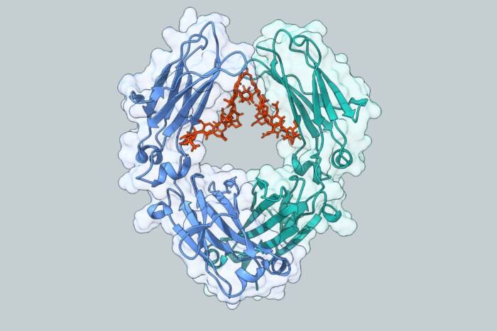
To accomplish this task, called exocytosis, the cell begins by sending newborn proteins to an organelle called the endoplasmic reticulum. Rockefeller Professor Günter Blobel, M.D., Ph.D., was awarded the Nobel Prize in 1999 for his demonstration of the zip codes that target nascent proteins to this organelle. Blobel, together with Simon, also showed that these zip codes act like keys to open water-filled pores that allow the proteins to enter this compartment.
Once inside the endoplasmic reticulum, protein exports are packaged into membrane-bound vesicles, which serve as personalized vehicles to transport the proteins to the surface. George Palade, Ph.D., an early pioneer in the field of cell biology at Rockefeller, was awarded the Nobel Prize in 1974 for his characterization of the pathway these vesicles take from the endoplasmic reticulum to the surface, where they finally dock with the membrane and release their contents.
But just how these protein-filled vesicles make their way to the cellular surface has remained unclear. While it is known that the vesicles are initially loaded onto the thick microtubules, it is not obvious where their journey ends. The most popular theory holds that microtubules make up the beginning of the trip, while actin filaments form the end.
Both microtubules and actin filaments constitute part of a cell’s cytoskeleton — a dynamic structure consisting of long fibrous proteins that provide both structural support and the ability to move.
Shining light on the problem
To address this issue from a new perspective, the Rockefeller researchers used Total Internal Reflection Fluorescence Microscopy (TIRFM). Based on an idea originally developed by Joseph Tyndall in 1860 — the same scientist who first explained why the sky is blue — TIRFM allows scientists to view the surface areas of living cells in real-time. By fluorescently tagging specific proteins, the researchers can observe the second-by-second happenings of individual proteins within their natural environment of the cell.
“As more and more proteins continue to be characterized, it is becoming increasing important to observe their normal behavior in cells,” says Simon. “We can now do this with TIRFM.”
There are two main types of microscopy: light and electron. While electron microscopy can achieve a finer spatial resolution than light microscopy, it does so at the expense of losing information about time; only static snapshots of cells can be taken using this technique. In contrast light microscopy allows cells and their components to be followed continuously through time.
Cellular secrets observed
The first surprise to strike the researchers came after fluorescently labeling the microtubules in the cell. What they saw through the microscope was a vast network of microtubules actively swimming about the surface layer of the cell. Previously, it was believed that microtubules functioned mainly at the heart of the cell and not near its surface.
The second surprise came after fluorescently tagging both actin filaments and microtubules, as well as a specific protein commonly exported from cells. This time the researchers observed slug-shaped vesicles filled with proteins wind their away through the cell, fuse with the membrane and release their contents to the outside. Their route? The vesicles followed only microtubules all the way out to the cellular membrane. Actin filaments played no role.
This finding was confirmed using inhibitors of both actin filaments and microtubules: only inhibitors of microtubules affected protein export, thereby proving their essential role.
Now that the researchers have the ability to look directly into the inner workings of the cell, they hope to address other important questions regarding exocytosis.
“How do microtubules contribute to the fusion of the vesicles? When and how are the components of this fusion machinery delivered to the appropriate sites?” asks Simon. “We hope to answer these and other questions by simply observing these components at work in their natural setting of the cell.”