Rockefeller researchers characterize yeast nuclear pore complex
 Researchers from The Rockefeller University and the University of Alberta in Canada have obtained the first comprehensive inventory of the protein components of the nuclear pore complex (NPC), an essential cellular structure that regulates transport between the nucleus and the cytoplasm. Along with the inventory, the researchers have suggested a simple model to explain how this multiptotein complex regulates macromolecular traffic into and out of the nucleus.
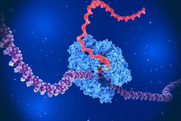
As the sole points of passage between the nucleus and the cytoplasm for macromolecules such as proteins, nuclear pore complexes form gateways that regulate the import and export of cargo to the nucleus. Cylinder-shaped and embedded in the nuclear envelope, NPCs determine what macromolecules are allowed to cross and ensure that the cargo proceeds in the right direction.
The NPC is one of the largest multiprotein complexes in the cell. Because of its size, it was thought that as many as 200 proteins, called nucleoporins or nups, would be found to contribute to its architecture. (By comparison, the ribosome, which directs protein synthesis, is one-twentieth the size of the NPC and comprises about 80 different proteins.) Recent research published in the Feb. 21 issue of the Journal of Cell Biology by two Rockefeller University laboratories and a laboratory at the University of Alberta in Canada, provides significant insights into this important organelle.

With a list of nucleoporins and their locations in the structure, Michael Rout and his colleagues set forth to understand how the NPC regulates transport across the nuclear envelope. Because the NPC must be both selective (discriminating what cargo can pass and what cannot) and directional (once something crosses the barrier it must stay there) it was thought some type of mechanochemical gating mechanism was at play. But they found no obvious motor proteins to support that theory. Instead, the team proposes a "virtual gate," called a Brownian affinity gate, to explain how the NPC regulates protein traffic into and out of the nucleus. Cargo cannot pass through the NPC unescorted; it must be attached to a transport factor. The researchers suggest proteins that do not bind the NPC can only transit the NPC by the unfavorable process of diffusion through the constricted central channel; this will bar most proteins from passage. However, transport factor-cargo complexes that specifically dock the NPC can offset this with the energetically favorable process of binding. This promotes the specific diffusional exchange of transport factors – laden with cargo – on and between the nuclear and cytoplasmic sides of the NPC through the central channel. The NPC is thus a "virtual gate" – because proteins that can bind the NPC pass the diffusion barrier of the central channel much more freely than those that do not, gating selectivity is achieved without necessarily invoking a gate composed of any moving parts. "Binding to the NPC is essential," says Graduate Fellow Tari Suprapto. "Once the transport factor-cargo complex binds to it, the complex can access both sides of the nuclear envelope because binding sites are found on both sides." The binding sites are symmetrically localized, says Suprapto, meaning that they are located on either side of the nuclear envelope in a similar distribution. Further out from the nuclear envelope are asymmetrically localized binding sites, or sites that are found on one side or the other, but not both. These asymmetric binding sites provide a target for transport factors to aim at in the compartment – nucleus or cytoplasm – they need to go to. But how do the transport factors know which is the correct target compartment to dump their cargo? Previous work from many groups, including the Blobel lab, showed the molecular environment in the target compartment promotes the termination of transport. An enzyme called Ran provides the directionality, telling the transport factors where they are. Ran exists in two forms. Inside the nucleus, Ran is bound to a nucleotide called GTP. When a cargo-laden transport factor reaches the nuclear side of the NPC during nuclear import, it is exposed to Ran-GTP, which promotes break up of the complex. On the cytoplasmic side of the NPC, Ran-GTPis converted to Ran-GDP, which promotes complexes to break up there, releasing the cargo in the right place. The model still needs to be tested, says Suprapto, and more work needs to be done to determine the molecular architecture. But, she says, "we have a whole battery of techniques that we can use to answer these questions."
In a commentary in the journal Nature, RU Professor Günter Blobel and Richard Wozniak write that “this work will point the way towards unveiling the secrets of other giant molecular machines… Not only does the work of Rout and his collaborators provide us with a vast amount of information on the molecular organization of the nuclear pore complex, it also gives us an inkling of the future of cell biology… Cell biologists can now enjoy the riches of our post-genomic era.”
About four years ago, while a research associate in Blobel’s lab, Michael Rout, now an assistant professor and head of the Laboratory of Cellular and Structural Biology, embarked on a project to obtain a comprehensive inventory of the proteins in the NPC to better understand how the NPC functions in transport across the nuclear envelope. He chose an approach that he had used to study another organelle called the spindle pole body (SPB) when he was a graduate student at the MRC Laboratory of Molecular Biology in Cambridge. Rout developed methods to isolate SPBs, and he generated antibodies that recognized and helped him to identify several distinct protein components in the SPB.
Rather than starting from single components of the NPC and working outward, the team sought to identify and characterize all the components together. This then allowed them to produce a comprehensive picture of the nuclear pore complex.
“Whenever you isolate a complex structure such as the NPC, a lot of other proteins, as well as nucleoporins, come along for the ride,” says Rout. A major problem the team had to confront was sorting through these proteins to find the real NPC components.
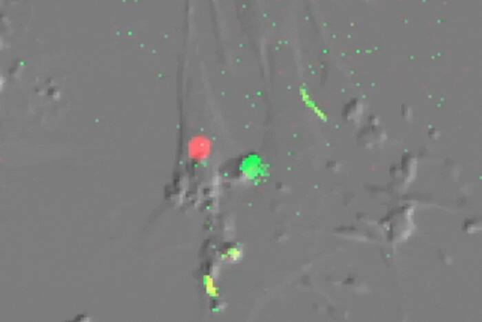
To characterize the NPC, Rout and his colleagues took yeast cells, separated the nuclei from the cytoplasm and then removed the NPCs from the nuclear envelope to produce an NPC-enriched solution. Working with Professor Brian Chait’s lab, Rout used a variety of chromatographic and mass spectrometric methods to identify 30 genuine NPCproteins from the hundreds of proteins that were present in their preparation.
The average mass of the nucleoporins is quite large, about four times the size of a ribosomal protein, and unlike ribosomal proteins, they exist in multiple copies in each NPC.
“It was somewhat surprising that so few proteins could make up a complex the size of an NPC,” says Tari Suprapto, a graduate fellow in Rout’s lab and one of the authors of the paper. “But if you take multiple copies of large proteins, it’s easily done.”
During the four-year-long project, three advances in the field occurred to dramatically speed up the identification and characterization of nucleoporins. First, the completion of the yeast genome project enabled the rapid mass spectrometric identification of yeast proteins.
Second, Rout and co-author John Aitchison, a former Blobel lab postdoctoral fellow who is now at the University of Alberta in Canada, developed a technique called genomic tagging to rapidly identify nucleoporins. Genomic tagging allows researchers to attach a molecular label to a protein, but in a way that doesn’t usually disrupt the normal expression of the gene that codes for the protein. This is important, says Suprapto, because any changes in protein synthesis would have skewed their results.
The third development was the refinement of mass spectrometric techniques by Professor Brian Chait and his Laboratory of Mass Spectrometry and Gaseous Ion Chemistry, whose team Rout credits with greatly decreasing the time required to identify proteins.
“We needed something that could give us an answer in days to hours,” says Rout. “That’s where Brian Chait’s fabulous technology came into play: the very rapid, very accurate identification of proteins, which he has been pioneering for years now.”
According to the researchers, the mass spectrometry techniques were 10 to 100 times more sensitive than they really needed, but this ensured that their identification of nucleoporins would be comprehensive.
Chait says this study stands out because it was among the first to rapidly and comprehensively identify all the components of a multiprotein complex, and it was done by six people working in three labs. In the past, he says, “something on this scale might have been considered virtually impossible.” Also, previous work on protein machines took decades or was not comprehensive. And completeness is of key importance, says Chait, to understanding function. “If you miss an essential component, you could get it all wrong.”
Chait says during the four years his lab worked with Rout, it continually improved and refined its mass spectrometric techniques. The technology, he says, is considerably better today than it was when they started.
“The real heroes are Mike and his lab,” Chait says. “It’s one thing to find a lot of proteins, but it’s quite another to identify them as part of a machine. They did an extraordinarily thorough study.”
In addition to Rout, Suprapto, Chait and Aitchison, other co-authors of the paper are graduate student Kelly Hjertaas from the University of Alberta and Yingming Zhao, a former graduate fellow in the Chait lab who now runs his own laboratory at the Mount Sinai School of Medicine. The research was funded in part by the National Institutes of Health, The Rockefeller University, the Howard Hughes Medical Institute, the Medical Research Council of Canada and the Alberta Heritage Foundation for Medical Research.