Our Research

Investigator, Howard Hughes Medical Institute
The Basics About Research in the Fuchs Laboratory
Why Use Skin as a Model System for Study?
As basic scientists with an interest in applying our knowledge to human medicine, we chose skin as a model system, because skin epithelium was the first and remains one of the few tissues of the body whose human and mouse stem cells (keratinocytes) can be maintained and propagated long-term in defined culture medium, without losing the ability to regenerate skin. This feature has been exploited for nearly 4 decades to treat burn patients with epidermal sheets generated from cultured stem cells. With gene therapy and Crispr/Cas technologies, devastating skin disorders can now be treated. The research opened similar doors for treating corneal blindness.
At the body surface, skin is readily accessible and particularly well-suited for mouse genetics and screens. After 25 years of performing classical knockouts and conditional knockouts to explore gene function, we developed methodology to perform gene manipulations specifically in the skin epithelium, but in a fraction of the time of conventional genetics. This revolutionary technology involving ultrasound-guided delivery to expose the early embryo to lentivirus in utero, now enables us to perform genome-wide screens to unearth the physiological relevance of genes in the skin of mice (Beronja et al., 2010). With this technology, we’ve performed screens to identify novel oncogenes, tumor-suppressors, translational regulators, cancer-causing microRNAs and more (Beronja et al., 2013; Schramek et al., 2014; Sendoel et al., 2017).
What kinds of questions does the laboratory address and what kinds of approaches do we take to address them?
We continue to expand upon our knowledge of skin stem cells. The global questions we are addressing center on how stem cells become mobilized from their niches to form tissues, how stem cells know when to stop making tissue after wound-repair and how these processes are deregulated in aging, inflammation and cancer. To do so requires a multifaceted approach to skin biology and the realization that there are many different cell types in the skin, many of which interact with stem cells and impact their behavior.
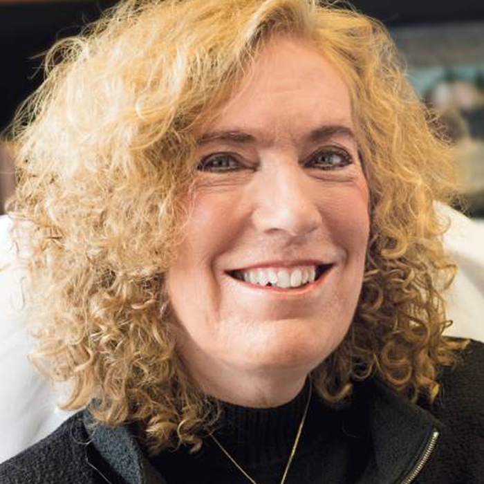
Elaine’s formal training is as a chemist, biochemist and cell biologist, but she focuses on questions that involve molecular, cell and developmental biology as well as mouse and human genetics. It’s not surprising to find that there is also a broad range of expertise and research approaches taken by individual lab members. The lab routinely uses high throughput technology and bioinformatics in their research.
Tissue fitness and stem cells. A long-standing focus of the lab has been on cytoskeletal and adhesion dynamics in morphogenesis. Postdocs and students in this area probe the regulation of actin, microtubule, intermediate filament dynamics and mechanical forces that operate during skin development and epithelial sheet formation. We also probe cytoskeletal interactions with integrins and cadherins in adhesion. We’ve engineered transgenic mice expressing fluorescently labeled cytoskeletal and adhesive proteins and use videomicroscopy, whole embryo imaging, mouse genetics and biochemistry, molecular and cell biology to study changes in cytoskeletal dynamics and adhesion in living cells and tissues. Such approaches led us to our discovery that mammalian epidermal cells stratify and differentiate by using a mechanism found in worms and flies to orient their mitotic spindle and divide asymmetrically (Lechler et al., 2005; Williams et al., 2014; Yang et al., 2017). We’ve also been interested in how stem cells are able to sense and eliminate unfit neighboring progenitors (Ellis et al., 2019). In doing so, we’ve unearthed a number of interesting findings relating to cell competition both during development and postnatally.
Understanding the molecular mechanisms involved in asymmetric divisions in stem cells, in changes in cytoskeletal-adhesive dynamics in epidermal migration and wound repair and in probing how stem cells eliminate unfit neighbors have been a focus of the lab’s spectrum, and ones with profound implications for cancer.
In the past decade, we also devised novel methods to mark, lineage-track and purify the epithelial stem cells from mouse skin, and we’ve employed RNAseq, single cell RNAseq and spatial transcriptomics technology (in collaboration with the Pe’er lab at MSKCC) to determine their global patterns of gene expression in vivo (Yang et al., 2017; Yuan et al., 2022; Niec et al., 2022). Many of these changes involve transcription factors and we’ve used conditional knockout technology to dissect their functions. We’re also employing in vivo landscaping for transcription factors, histone modifications, histone modifying enzymes and chromatin accessibility to understand how skin stem cells coordinate transcription factors and epigenetic modifiers to change their chromatin landscape as stem cells receive signals from their environment and transition from a non-tissue generating (quiescent) state to one where they are either making or repairing a tissue (H. Yang et al., 2017; Y. Yang et al., 2023). The hair follicle is ideal for these studies, since in the mouse, every HF has a stem cell niche, and all the HF stem cell niches act synchronously, either remaining in quiescence for weeks, or actively regrowing the HF and making hair.
Stem cells, Aging and the Niche. Quiescent HF stem cells are in a WNT-inhibited and BMP-rich environment and become activated to make upon directional WNT signaling and inhibition of BMP signaling. We’ve begun to dissect the downstream targets of these pathways, and have shown that while WNT inhibition represses stem cell fate specification, BMP signaling maintains quiescence by a mechanism that involves NFATc1, FOXc1 and ID transcription factors, downstream of canonical pSMAD1/SMAD4. Elevated BMP signaling also occurs in aging skin, providing a mechanism for why our hair becomes sparser as we age (Keyes et al., 2013; 2016; Ge et al., 2020 PNAS).
We’d like to know more about the molecular coordination of these and other signaling pathways that operate on skin stem cells, and how they elicit changes in chromatin and transcriptome dynamics. We want to elucidate the molecular cross-talk between epidermal/HF stem cells and other permanent or transient residents of the skin, including dermal fibroblasts, immune cells, blood vessels, sensory neurons and melanocytes. As we dissect the cross-talk that elicits dramatic changes in chromatin, transcriptional and translational landscapes in the skin stem cell niches, we also want to elucidate the significance of the myriad of downstream changes in proliferation, cell adhesion, cytoskeletal dynamics and cell polarity.